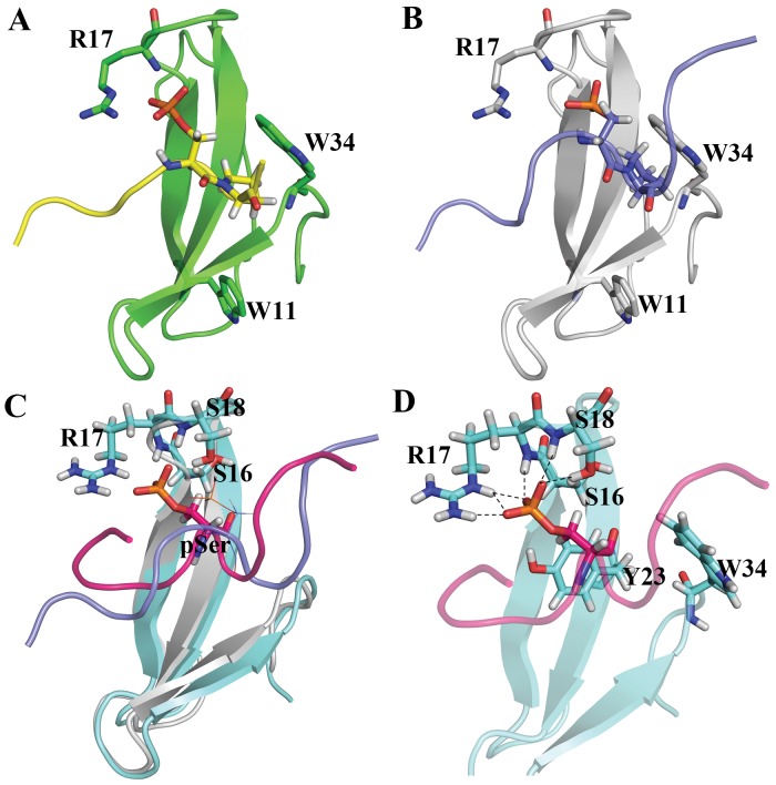
Figure 4. Model building and MD simulation result of Pin1-pS87 peptide.
(A) Crystal structure of the Pin1 WW domain with C-terminal domain (CTD) of RNA polymerase II phosphopeptide. (B) Initial model of Pin1 WW domain with pS87 Bcl-2 phosphopeptide. Both the tryptophan residues and the phosphoserine-interacting arginine residue are highlighted as sticks, and labeled accordingly. Both phosphopeptides are highlighted with yellow (CTD peptide) and blue (pS87 Bcl-2 peptide), and the pSer-Pro motif in both the phosphopeptides is represented as sticks. (C) Initial and representative frames of MD simulation. Protein is represented with gray and cyan; pS87 peptide is highlighted with purple and pink for the initial and final frames, respectively. (D) Hydrogen bond interaction patterns of the phosphoserine with the WW domain residues. All three residues (2 serines and 1 arginine) are highlighted with sticks and labeled. Hydrogen bonds between the phosphoserine and the WW domain residues are represented with dashed lines.

