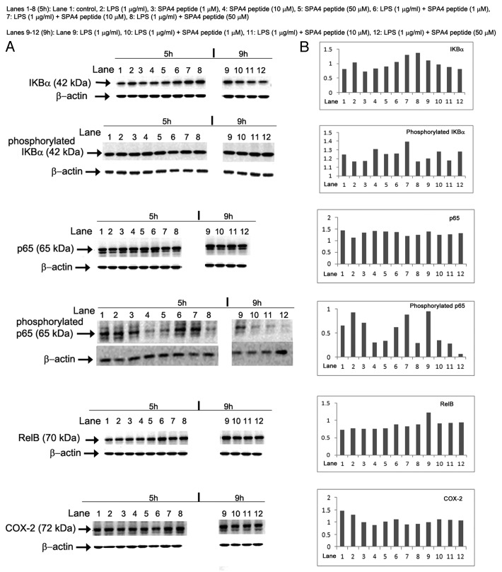
Figure 5. Effects of the SPA4 peptide on NFκB signal transduction. (A, B) Ten µg total proteins were run on 4–20% Tris-glycine SDS-PAGE gels in partially-reducing condition (heating at 95°C for 5 min, no reducing agent). Separated proteins were immunoblotted with specific antibodies. Images of immunoreactive bands were acquired and analyzed densitometrically. (A) Acquired images of immunoreactive bands of IKBα, phosphorylated IKBα, p65, phosphorylated p65, RelB and COX-2 in SW480 cells treated with lipopolysaccharide (LPS) ± SPA4 peptide for a total period of 5 h and 9 h (see Figure 4). (B) Densitometric readings of the indicated immunoreactive bands normalized to those of β-actin, which was employed as a loading control. Results are from one out of two independent experiments.

An official website of the United States government
Here's how you know
Official websites use .gov
A
.gov website belongs to an official
government organization in the United States.
Secure .gov websites use HTTPS
A lock (
) or https:// means you've safely
connected to the .gov website. Share sensitive
information only on official, secure websites.
