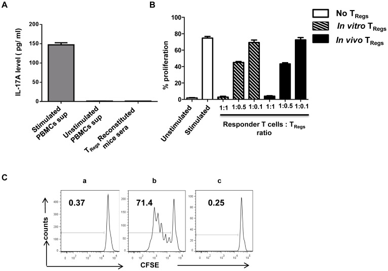
Figure 3. In vivo expanded TRegs do not convert to Th17 cells and retain their characteristic suppressive function.
(A) IL-17A levels in the sera of nTReg reconstituted animals were analyzed by ELISA. Error bars indicate standard deviation for six different animals tested. Supernatants from normal human PBMCs stimulated with PMA (50 ng/ml) and ionomycin (1 µg/ml) served as positive control. (B) TRegs obtained from reconstituted mice and in vitro expanded TRegs were tested for their ability to suppress proliferation of autologous CD4+CD25− T cells at different TReg to CD4+CD25− responder T cell ratios. Responder cells were stimulated with anti-CD3/CD28 beads and CFSE dilution was analyzed after 5 days of culture under the following conditions: autologous T cells cultured in medium without stimulation (unstimulated), stimulated with anti-CD3/anti-CD28 coated micro beads in the absence of TRegs (stimulated), in the presence of cultured TRegs (in vitro TRegs) or in the presence of TRegs isolated from reconstituted mice 12 days after reconstitution (In vivo TRegs). Composite data from all the three animals tested are shown. (C) The functional stability of TRegs obtained 33 days after in vivo reconstitution was also analyzed by CFSE dilution of CD4+CD25− responder cells. Representative data from one of two animals tested is shown. Data depicts CD4+CD25− responder cells cultured with medium alone (a), stimulated in the absence of in vivo TRegs (b) and stimulated in the presence of in vivo TRegs (c).

