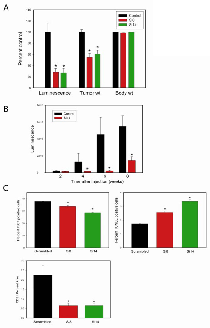
Figure 2. SiRNAs targeting the Type III T/E fusion mRNA inhibit tumor progression in vivo.
A. Nude mice were injected orthotopically with VCaP expressing luciferase. After 10 days mice were treated with DOPC liposomes containing scrambled (control) or targeted siRNAs (Si8 or Si14) twice weekly for 4 weeks (150 ug/kg). Luciferase flux of tumors prior to euthanasia and tumor and body weights at termination of treatment are shown. The control group contained 9 mice while each targeted group contained 11 mice. B. Nude mice were injected subcutaneously with VCaP expressing luciferase (time 0). After two weeks mice were treated with DOPC liposomes containing scrambled (control) or targeted Si14 twice weekly for 6 weeks. Luciferase flux of tumors at indicated time is shown. The control group had 7 mice and the Si14 treated group had 8 mice. C. Mean percent nuclei stained with Ki67 or TUNEL or mean tumor area stained with anti-CD31 in treated and control tumors. Values are mean +/− SEM normalized to control (100%). Asterisks indicate statistically significant differences between targeted siRNA and scrambled control.

