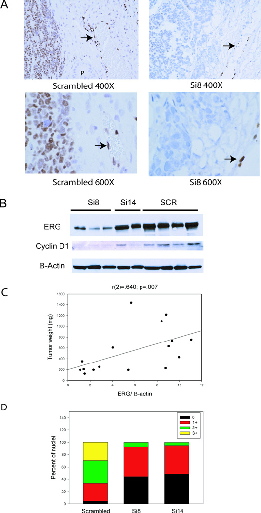
Figure 3. Knockdown of ERG protein in tumors treated with targeted siRNAs.
A. Immunohistochemistry with anti-ERG antibody of orthotopic VCaP tumors treated with scrambled control siRNA or Si8. VCaP tumors are to the left; blood vessels adjacent to tumors with endothelial cells expressing ERG are indicated by arrows. A residual normal gland is marked with “P” in the scrambled control (400X). Original magnification: 400X or 600X. B. Representative Western blot of VCaP tumor extracts with antibodies against ERG or Cyclin D1. β-actin is a loading control. C. Correlation between ERG protein level by quantitative Western blotting, normalized to β-actin, and final tumor weight. D. Percentage of nuclei expressing the ERG target NFκB p65 phospho-Ser536 as evaluated my Inform image analysis in tumors treated with control or targeted SiRNAs. Asterisks indicate statistically significant differences in the percentage of moderate and strong (2+ and 3+) between control and targeted groups by t-test (p<.001).

