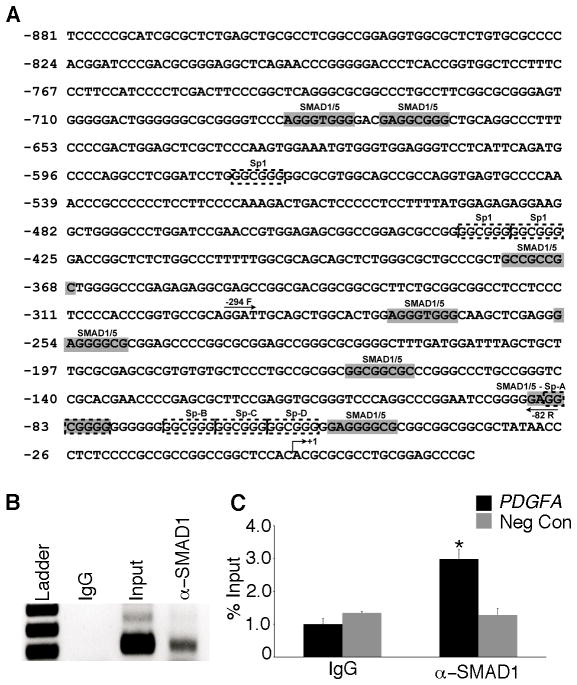
Figure 5.
SMAD1/5 bind to the PDGFA promoter. (A) In silico promoter analysis identified several putative SMAD1/5 binding sites (gray shaded region) in the human PDGFA promoter region. Also shown are binding sites for Sp1 (dashed boxes), a known positive regulator for PDGFA promoter activity. The arrows in the promoter region indicate the sites where forward (-294F) and reverse (-82R) primers are designed for the ChIP assay. (B) Chromatin immunoprecipitation (ChIP) assay with an anti-SMAD1/5 antibody or control IgG was performed on cell extracts from COV434 cells demonstrated the in vivo binding of SMAD1/5 to the PDGFA promoter. (C) Real-time PCR analysis of immunoprecipitated DNA using locus specific primers demonstrated three-fold enrichment (ChIP/Input DNA) of SMAD1/5 occupancy at the PDGFA promoter compared to negative control (no locus-specific primers) and IgG. Asterisks indicate statistical significance (n=3; P< 0.05).

