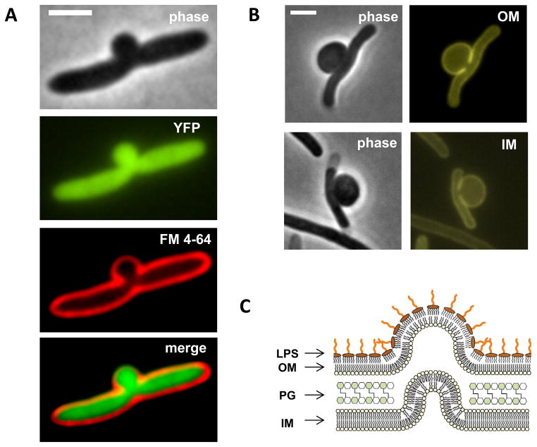
Figure 1. Both inner and outer membranes are intact in bulging cells.
(A, B) Phase contrast and fluorescence images of bulging cells under cephalexin treatment (A, cytoplasmic YFP and FM 4–64 membrane staining; B, ZipA-mCherry, IM marker; ssPal-mCherry, OM marker). Membrane protrusions indicate PG defects at the potential division site. Leakage of cytoplasmic YFP into the bulge suggests redistribution of cytosol materials from cell filament to the midcell bulge. (C) Schematic illustration of a bulge as a cell wall-less cytosolic protrusion surrounded by both IM and OM. LPS is exclusively located at the outer leaflet of the OM and its long glycan chains facilitate a micelle-like close packing structure. All scale bars represent 2 μm. See also Figure S1.

