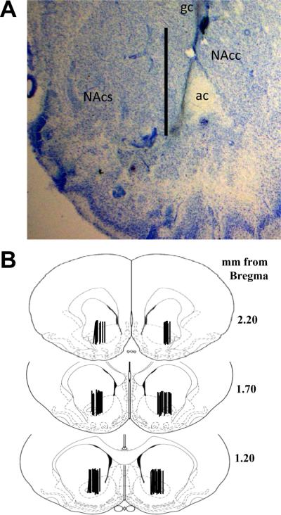
Figure 5.
The placement of the microdialysis probes resulted in sampling primarily from the nucleus accumbens core, although aspects of ventral nucleus accumbens shell and the striatum dorsal to the nucleus accumbens were sampled as well. (a) A representative coronal section illustrating the placement of microdialysis probes. The tract created extends approximately 3 mm from the guide cannula with sampling only occurring from the most ventral 2 mm because this is the portion of the probe containing the dialysis membrane. Scale bar = 2 mm, ac (anterior commissure), NAcc (nucleus accumbens core), NAcs (nucleus accumbens shell). (b) Schematics depicting the distribution of the active membrane portion of the microdialysis probes illustrate the regions sampled in this study.

