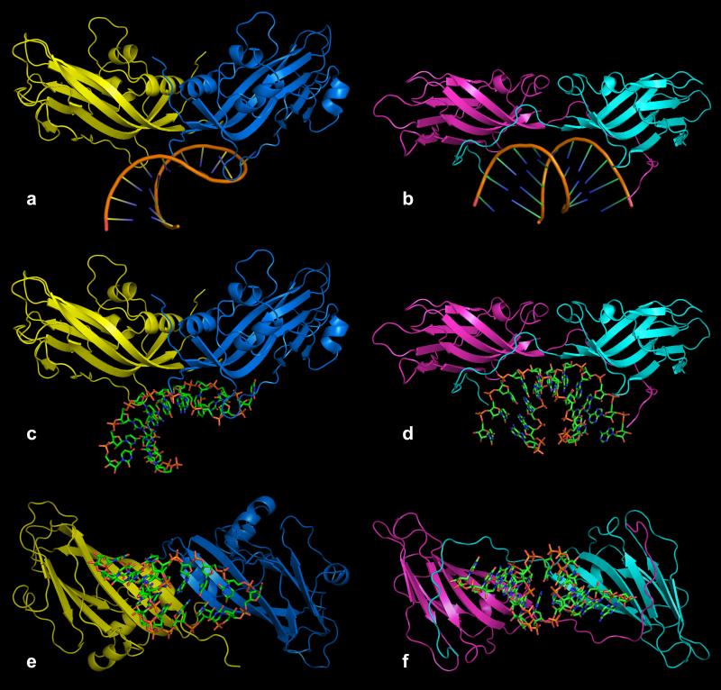
Fig. 10.
Comparison of the capsid dimer-RNA complex of PMV with the equivalent dimer-RNA complex of STMV. In (a), (c) and (e) are ribbon representations of A (blue) and B (yellow) subunits of PMV (related by an icosahedral quasi twofold axis) and their bound RNA. In (b), (d) and (f) are comparable views of the capsid dimer-RNA complex from STMV. Panels (a-d) are views perpendicular to the twofold axes and panels (e) and (f) are views along the twofold axes from the particle centers.

