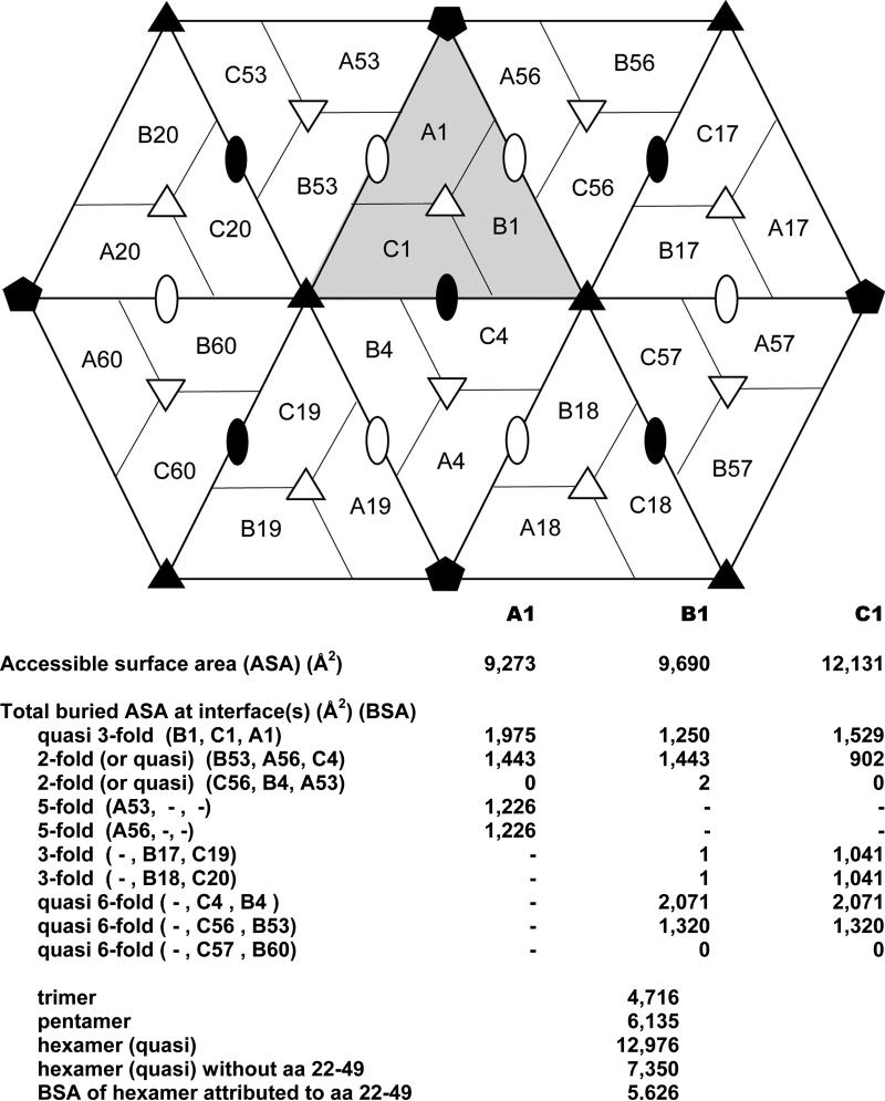
Fig. 4.
Accessible and buried surface areas of subunits in the PMV capsid. The diagram shows a primary trimer, labeled as A1, B1, and C1, and the neighboring subunits. Exact icosahedral symmetry axes are indicated by solid symbols and quasi symmetry axes by open symbols. Below the diagram, accessible and buried surface areas are tabulated for each subunit of the primary trimer for each indicated relationship and total buried surface areas are given for subunit assemblies.

