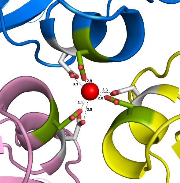
Fig. 8.
The putative calcium ion on the quasi threefold axis is shown as a red sphere, with its six octahedrally arranged carboxylate ligands forming a cage about it. Three of the side chains are provided by symmetry related Asp198 (green) and three by symmetry related Asp201 (white). These were the only calcium ions observed in the PMV capsid. The distances are for the primary quasi trimer A1, B1, and C1 of particle 1.

