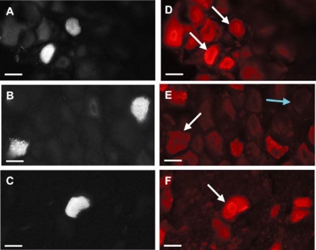
Figure 1.

PAR2 expression in knee joint afferent neuronal cell bodies. Left: Retrograde labelling of knee joint afferent DRGs with Fluoro-Gold taken from L3 (A), L4 (B) and L5 (C). Right: Immunofluorescent imaging using Cy3 reveals a co-localization of PAR2-positive cells in the corresponding L3–L5 DRGs (D–F). Scale bar, 30 μm.
