
Figure 5.
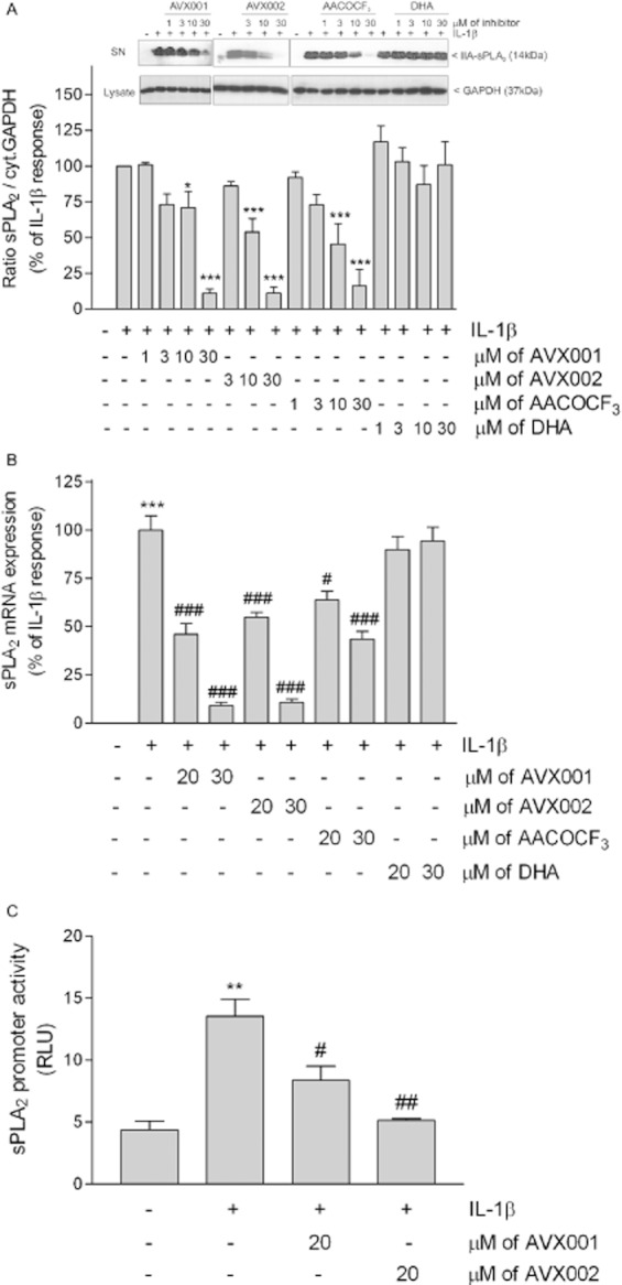
Effect of AVX001 and AVX002 on IL-1β-stimulated sPLA2 protein (A), mRNA expression (B) and promoter activity (C) in mesangial cells. Quiescent cells were stimulated with either DMEM (-), or IL-1β (1 nM) in the absence (-) or presence of the indicated concentrations of AVX001, AVX002, AACOCF3 and DHA (all inhibitors were added as a pretreatment for 90 min before stimulation). (A) Supernatants were taken for protein precipitation, separated by SDS-PAGE, transfered to membranes and subjected to Western blot analysis using a monoclonal antibody against rat sPLA2. Corresponding cell lysates were stained for GAPDH and used for normalization. Bands were densitometrically evaluated and the ratio between secreted sPLA2 and cytosolic GAPDH was calculated. Results are expressed as % of IL-1β stimulation. Data are means ± SD (n = 3–5). The inset shows representative Western blots of sPLA2 (upper panels) from supernatants (SN) and GAPDH (lower panels) from cell lysates. (B) Cells were taken for RNA extraction and subjected to quantitative PCR analysis of rat IIA-sPLA2 and 18S RNA. ΔΔCt values were calculated and results are expressed as % of maximal IL-1β-stimulated response and are means ± SD (n = 3–5). (C) Cells were transfected with the sPLA2 promoter construct plus a plasmid coding for Renilla luciferase. After transfection, cells were stimulated for 24 h with vehicle (-), IL-1β (1 nM), or IL-1β in the absence or presence of AVX001 or AVX002 (both at 20 μM). Both inhibitors were added as a pretreatment 90 min before stimulation. sPLA2 promoter activity was calculated and results are expressed as relative luciferase units (RLU) and are means ± SD (n = 3). **P < 0.01, ***P < 0.001 considered statistically significant when compared with the control values; #P < 0.05, ##P < 0.01, ###P < 0.001 when compared with the IL-1β-stimulated values.