. 2012 Dec 18;1:e00117. doi: 10.7554/eLife.00117

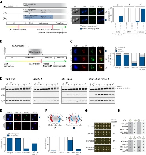
Figure 5. Transient disruption of microtubule–kinetochore interactions suppresses the chromosome segregation defects in CUP-CLB3 cells.

(A) Wild-type (A10684) and GAL-CDC5 GAL-MAM1 (A26546) cells, carrying a MET-CDC20 allele and CENIV-GFP dots, were monitored for chromosome segregation in anaphase (see ‘Materials and methods’ for details). MT = microtubule, KT = kinetochore, (n = 100). The fraction of anaphase cells that segregate or cosegregate sister chromatids was compared between GAL-CDC5 GAL-MAM1 condition (2) and GAL-CDC5 GAL-MAM1 condition (3) using a chi-square test (df 1) χ2 = 59.71, p<0.0001. (B) Schematic description of the experimental regime used for (C) through (H) see ‘Materials and methods’ for details. (C) Localization of Lrs4-13myc (green) in mononucleates relative to Ndc10-6HA (red) determined by nuclear spreads (n > 40) and (D) phosphorylation of Lrs4-13myc determined by gel mobility shift in wild-type (A29612), ndc80-1 (A29614), CUP-CLB3 (A29616) and CUP-CLB3 ndc80-1 (A29618). For (C), using a chi-square test (df 2) the fraction of spread nuclei that display colocalized, partial or mislocalized Lrs4 with respect to Ndc10 was compared between wild-type and ndc80-1 χ2 = 0.9668, p=0.6167 and between CUP-CLB3 and CUP-CLB3 ndc80-1 χ2 = 56.34, p<0.0001. (E) Localization of Rec8-13myc (green) in binucleates relative to Ndc10-6HA (red) determined by nuclear spreads in wild-type (A28716), ndc80-1 (A28720), CUP-CLB3 (A28718) and CUP-CLB3 ndc80-1 (A28722) (n > 40). Using a chi-square test (df 1) the fraction of spread nuclei that were Rec8 positive or negative was compared between wild-type and ndc80-1 χ2 = 1.185, p=0.2764 and between CUP-CLB3 and CUP-CLB3 ndc80-1 χ2 = 23.96, p<0.0001. (F) Segregation of sister chromatids using heterozygous CENV-GFP dots quantified in binucleates (n = 100) and (G) spore viability from wild-type (A22678), ndc80-1 (A28621), CUP-CLB3 (A22702) and CUP-CLB3 ndc80-1 (A28623) (n = 40 tetrads for wild-type and ndc80-1, n > 60 tetrads for CUP-CLB3 and CUP-CLB3 ndc80-1) (nonpermissive temperature >36°C). Using a chi-square test (df 1) the fraction of binucleates with a reductional or equational division was compared between CUP-CLB3 and CUP-CLB3 ndc80-1 χ2 = 24.18, p<0.0001. (G) Segregation of chromosome V using homozygous CENV-GFP dots quantified in tetranucleates from wild-type (A22688), ndc80-1 (A28625), CUP-CLB3 (A22708) and CUP-CLB3 ndc80-1 (A28627). Top panel: cells kept at 25°C for the duration of the experiment. Bottom panel: Cells treated as in (B) but monitored after meiosis II (n = 100).

DOI: http://dx.doi.org/10.7554/eLife.00117.029

Figure 5.

Figure 5—figure supplement 1. Sporulation efficiency of ndc80-1 mutants.

Figure 5—figure supplement 1.

Wild-type (A22678) and ndc80-1 (A28221) cells were induced to sporulate at 25°C. 2 hr 30 min after transfer into sporulation medium, cells were shifted to the indicated temperature and sporulation efficiency was determined after 24 hr.
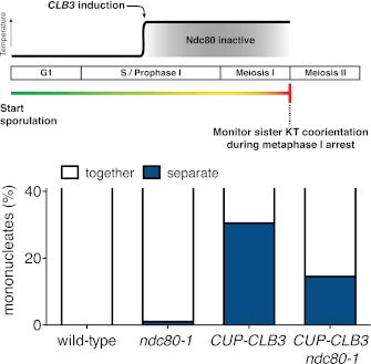
Figure 5—figure supplement 2. Sister kinetochore coorientation in ndc80-1 cells under a continuous inactivation regime at 34°C during a metaphase I arrest.

Figure 5—figure supplement 2.

Wild-type (A7118), CUP-CLB3 (A23074), ndc80-1 (A29690) and ndc80-1 CUP-CLB3 (A29692) cells also carrying the cdc20-mn allele, were induced to sporulate at 25°C. 2 hr 45 min after transfer into sporulation medium, CuSO4 (50 μM) was added and concurrently, cultures were shifted to 34°C. The percentage of mononucleate cells with separated CENV-GFP dots was determined 7 hr 30 min after transfer into sporulation medium when cells were arrested in metaphase I (n = 100). The fraction of nuclei that display sister kinetochores as separate or together was compared between CUP-CLB3 and CUP-CLB3 ndc80-1 using a chi-square test (df 1) χ2 = 7.228, p=0.0072.
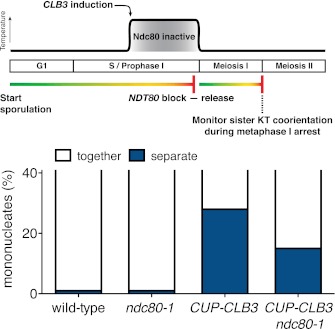
Figure 5—figure supplement 3. Sister kinetochore coorientation in ndc80-1 cells after transient inactivation regime at 34°C during a metaphase I arrest.

Figure 5—figure supplement 3.

Wild-type (A20958), CUP-CLB3 (A23076), ndc80-1 (A29718) and ndc80-1 CUP-CLB3 (A29720) cells also carrying the GAL4-ER and GAL-NDT80 fusions and the cdc20-mn allele were induced to sporulate at 25°C. 2 hr 45 min after transfer into sporulation medium CuSO4 (50 μM) was added and concurrently, cultures were shifted to 34°C. After 5 hr, when cells had arrested in the NDT80 arrest, cells were released from the NDT80 block and transferred to 25°C. The percentage of mononucleate cells with separated CENV-GFP dots was determined 7 hr 30 min after transfer into sporulation medium when cells were arrested in metaphase I (n = 100). The fraction of nuclei that display sister kinetochores as separate or together was compared between CUP-CLB3 and CUP-CLB3 ndc80-1 using a chi-square test (df 1) χ2 = 5.007, p=0.0252.
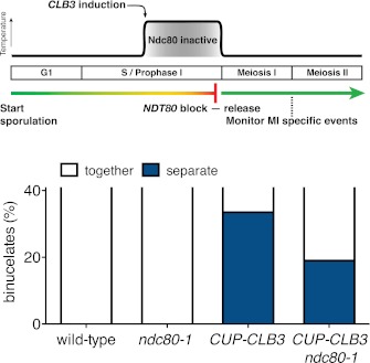
Figure 5—figure supplement 4. Meiosis I chromosome segregation in ndc80-1 cells after a transient inactivation regime at 34°C.

Figure 5—figure supplement 4.

Wild-type (A22678), ndc80-1 (A28621), CUP-CLB3 (A22702) and CUP-CLB3 ndc80-1 (A28623) cells also carrying the GAL4-ER and GAL-NDT80 fusions were induced to sporulate at 25°C. 2 hr 45 min after transfer into sporulation medium CuSO4 (50 μM) was added and concurrently, cultures were shifted to 34°C. After 5 hr, when cells had arrested in the NDT80 block, cells were released and transferred to 25°C. The percentage of binucleate cells with segregated heterozygous CENV-GFP dots was determined 7 hr 30 min after transfer into sporulation medium (n = 100). Note that a greater suppression of meiosis I sister chromatid segregation was observed in ndc80-1 CUP-CLB3 cells when cells were incubated at temperatures higher than 34°C (Figure 5F and data not shown). The fraction of binucleates that underwent reductional or equational division was compared between CUP-CLB3 and CUP-CLB3 ndc80-1 using a chi-square test (df 1) χ2 = 5.776, p=0.0162.
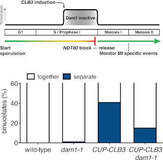
Figure 5—figure supplement 5. Transient disruption of microtubule–kinetochore interactions using dam1-1 allele restores meiosis I chromosome segregation in CUP-CLB3 cells.

Figure 5—figure supplement 5.

Wild-type (A22678), dam1-1 (A28311), CUP-CLB3 (A22702) and CUP-CLB3 dam1-1 (A28341) cells also carrying the GAL4-ER and GAL-NDT80 fusions were induced to sporulate at 25°C. 2 hr 45 min after transfer into sporulation medium CuSO4 (50 μM) was added and concurrently, cultures were shifted to 34°C. After 5 hr, when cells had arrested in the NDT80 block, cells were released and transferred to 25°C. The percentage of binucleate cells with segregated heterozygous CENV-GFP dots was determined 7 hr 30 min after transfer into sporulation medium (n = 100). The fraction of binucleates that underwent reductional or equational division was compared between CUP-CLB3 and CUP-CLB3 dam1-1 using a chi-square test (df 1) χ2 = 16.77, p<0.0001.
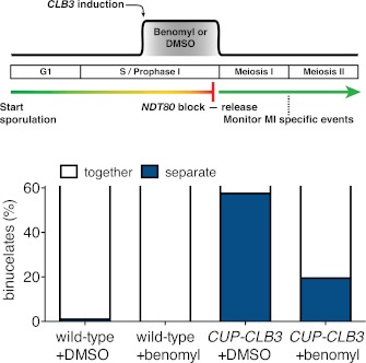
Figure 5—figure supplement 6. Transient disruption of microtubule–kinetochore interactions by benomyl treatment restores meiosis I chromosome segregation in CUP-CLB3 cells.

Figure 5—figure supplement 6.

Wild-type (A22678) and CUP-CLB3 (A22702) cells also carrying the GAL4-ER and GAL-NDT80 fusions were induced to sporulate at 30°C. 2 hr 15 min after transfer into sporulation medium CuSO4 (50 μM) was added and concurrently, cells were treated with DMSO or benomyl (120 μg/ml). Cells were subsequently released from NDT80 block 4 hr 30 min after transfer into sporulation medium and benomyl was washed out concomitant with NDT80-block release. The percentage of binucleate cells with segregated heterozygous CENV-GFP dots was determined 6 hr after transfer into sporulation medium (n = 100). See ‘Materials and methods’ for further details. The fraction of binucleates that underwent reductional or equational division was compared between CUP-CLB3 + DMSO and CUP-CLB3 + benomyl using a chi-square test (df 1) χ2 = 32.12, p<0.0001.
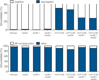
Figure 5—figure supplement 7. Transient disruption of microtubule–kinetochore interactions during S phase/prophase I suppresses CUP-CLB3-induced meiosis I sister chromatid segregation in a spindle assembly checkpoint independent manner.

Figure 5—figure supplement 7.

(Top panel) wild-type (A22678), mad3∆ (A30386), ndc80-1 (A28621), ndc80-1 mad3∆ (A30390), CUP-CLB3 (A22702), CUP-CLB3 mad3∆ (A30388), CUP-CLB3 ndc80-1 (A28623) and CUP-CLB3 ndc80-1 mad3∆ (A30392) cells also carrying the GAL4-ER and GAL-NDT80 fusions were induced to sporulate at 25°C. 2 hr 45 min after transfer into sporulation medium CuSO4 (50 μM) was added and concurrently, cultures were shifted to 36°C. Cells were subsequently released from NDT80 block at 5 hr and transferred to 25°C. Percent binucleates with segregated heterozygous CENV-GFP dots was determined (n = 100). (Bottom panel) wild-type (A22688), mad3∆ (A30638), ndc80-1 (A28625), ndc80-1 mad3∆ (A30642), CUP-CLB3 (A22708), CUP-CLB3 mad3∆ (A30640), CUP-CLB3 ndc80-1 (A28627) and CUP-CLB3 ndc80-1 mad3∆ (A30644) cells also carrying the GAL4-ER and GAL-NDT80 fusions were induced to sporulate at 25°C. 2 hr 45 min after transfer to sporulation medium CuSO4 (50 μM) was added and concurrently, cultures were shifted to 36°C. After 5 hr, when cells had arrested in the NDT80 block, cells were released and transferred to 25°C. The percentage of binucleate cells with segregated homozygous CENV-GFP dots was determined 7 hr 30 min after transfer into sporulation medium. Binucleate cells with GFP signal in only one of the two nuclei were categorized as having experienced a meiosis I non-disjunction event (n = 100). For top panel, using a chi-square test (df 1) the fraction of binucleates that underwent reductional or equational division was compared between CUP-CLB3 and CUP-CLB3 mad3∆ χ2 = 0.1800, p=0.6714 and between CUP-CLB3 ndc80-1 and CUP-CLB3 ndc80-1 mad3∆ χ2 = 0.02454, p=0.8755. For bottom panel, using a chi-square test (df 1) the fraction of binucleates that displayed MI nondisjunction or other was compared between CUP-CLB3 and CUP-CLB3 mad3∆ χ2 = 1.228, p=0.2678 and between CUP-CLB3 ndc80-1 and CUP-CLB3 ndc80-1 mad3∆ χ2 = 0.6486, p=0.4206.
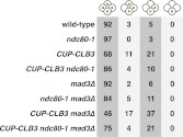
Figure 5—figure supplement 8. Transient disruption of microtubule–kinetochore interactions during S phase/prophase I restores meiotic chromosome segregation in CUP-CLB3 cells in a spindle assembly checkpoint independent manner.

Figure 5—figure supplement 8.

Wild-type (A22688), ndc80-1 (A28625), CUP-CLB3 (A22708), CUP-CLB3 ndc80-1 (A28627), mad3∆ (A30638), ndc80-1 mad3∆ (A30642), CUP-CLB3 mad3∆ (A30640) and CUP-CLB3 ndc80-1 mad3∆ (A30644) cells also carrying the GAL4-ER and GAL-NDT80 fusions were induced to sporulate at 25°C. 2 hr 45 min after transfer to sporulation medium CuSO4 (50 μM) was added and concurrently, cultures were shifted to 36°C. After 5 hr, when cells had arrested in the NDT80 block, cells were released and transferred to 25°C. Segregation of homozygous CENV-GFP dots was determined in tetranucleates 12 hr after transfer into sporulation medium (n = 100).
Figure 5—figure supplement 9. Transient ndc80-1 inactivation does not alter in vitro Cdk1 activity.

Figure 5—figure supplement 9.

Wild-type (A25508), ndc80-1 (A33203), CUP-CLB3 (A33201) and CUP-CLB3 ndc80-1 (A33205) cells also carrying the GAL4-ER and GAL-NDT80 fusions were induced to sporulate at 25°C. 2 hr 45 min after transfer to sporulation medium CuSO4 (50 μM) was added and concurrently, cultures were shifted to 35°C. Samples were harvested 5 hr post transfer to sporulation medium, when cells were arrested in the NDT80 block. In vitro kinase assays were performed with Cdc28-3V5 (Cdk1) immunoprecipitated from prophase I-arrested samples. Amounts of phosphorylated Histone H1 and immunoprecipitated Cdc28-3V5 are shown.