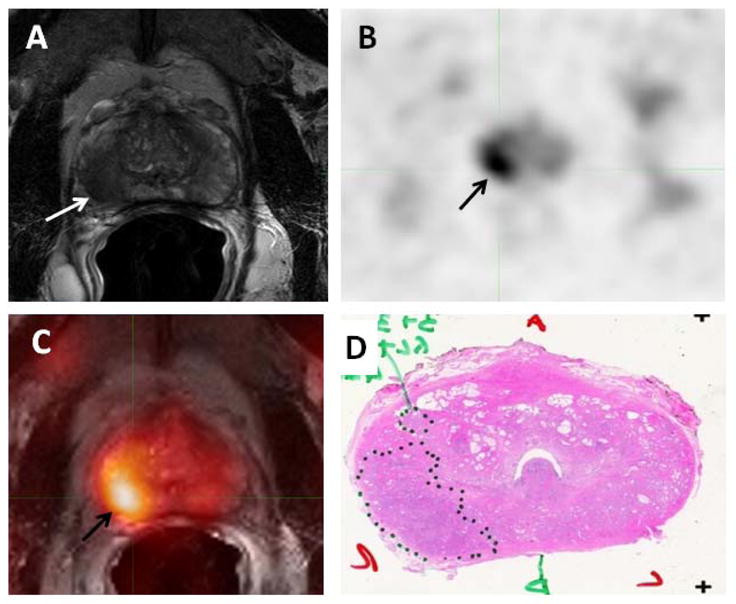
Figure 2.

58-year-old male with prostate cancer. Axial T2-weighted MRI (A) demonstrates a low signal intensity focus in the right mid gland peripheral zone (white arrow), which shows 11C-Acetate uptake in the axial PET image (black arrow) (B). Fused 11C-Acetate PET/MRI (C) better localizes the tumor (black arrow). Corresponding pathology shows a Gleason 3+4 tumor in the right mid-peripheral zone (inked in black) (D).
