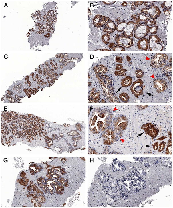
Figure 5.
Immunohistochemical staining of fatty acid synthase in prostate cancers. Both 11C-acetate negative and positive cases were stained. Note the intense brown staining of most cells against the background stroma in most of the specimens, which can be recognized even at low power (A, C, E). Discrete differences in staining intensity were seen between benign tissue (arrowheads) and tumor (arrows); however no difference was seen between (A–D) 11C-acetate-negative cases and (E–G) 11C-acetate-positive cases. Negative controls showing no FAS staining (H). Magnification: (A, C, E)=5X; (B, D, F)=20X; G–H=10X.

