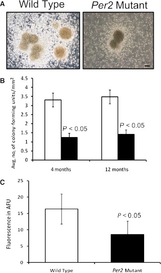
FIG. 6.
Decrease in proliferation and NO of Per2 mutant animals. A: 1 × 10⁴ mouse bone marrow mononuclear cells were placed on methylcellulose semisolid media under well-humidified conditions. After 7 days, colony-forming units were detected in Per2 mutant and wild-type mice. Representative images from colony-forming units of wild-type or Per2 mutant mice are shown. B: Quantification of colony-forming units showed a significant decrease in numbers of colony-forming units in Per2 mutant mice compared with wild-type control. C: BMPCs isolated from Per2 mutant mice showed a significant decrease in expression of NO as determined by DAF-FM fluorescence. n = 7 for 4-month-old mice; n = 3 for 12-month-old mice; white bars, wild type; black bars, Per2 mutant. AFU, arbitrary fluorescence unit. (A high-quality color representation of this figure is available in the online issue.)

