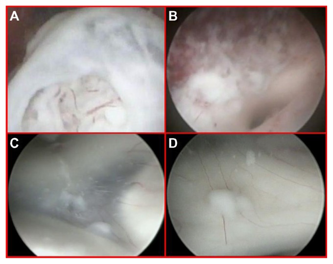
Figure 3.
Close-up endoscopic third ventricle views.
Notes: The nodule inside the third ventricle, just beneath the opened ependymal layer (A). Endoscopic view after tumor removal with the origin of the sylvian aqueduct (B). The floor of the third ventricle detecting a diffuse tumor infiltration composed of small adjacent nodules lying beneath the ependyma, unidentified by MRI and PET scan (C and D).

