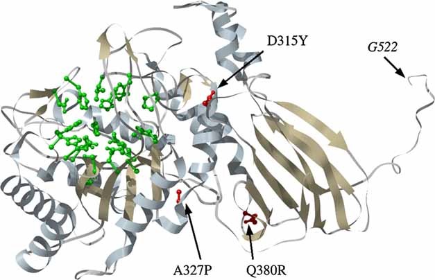
Fig 2.

Missense mutations in the 3D structure of the IDUA model of Rempel et al. 2005. The mutated residues are shown in red and the residues depicted in green are predicted to be within the active site. The model does not display the structure of the 130 amino acids at the C terminus (523–653). The last shown residue is G522 (for explanation please see the text). [Color figure can be viewed in the online issue, which is available at http://www.interscience.wiley.com.]
