Figure 4.
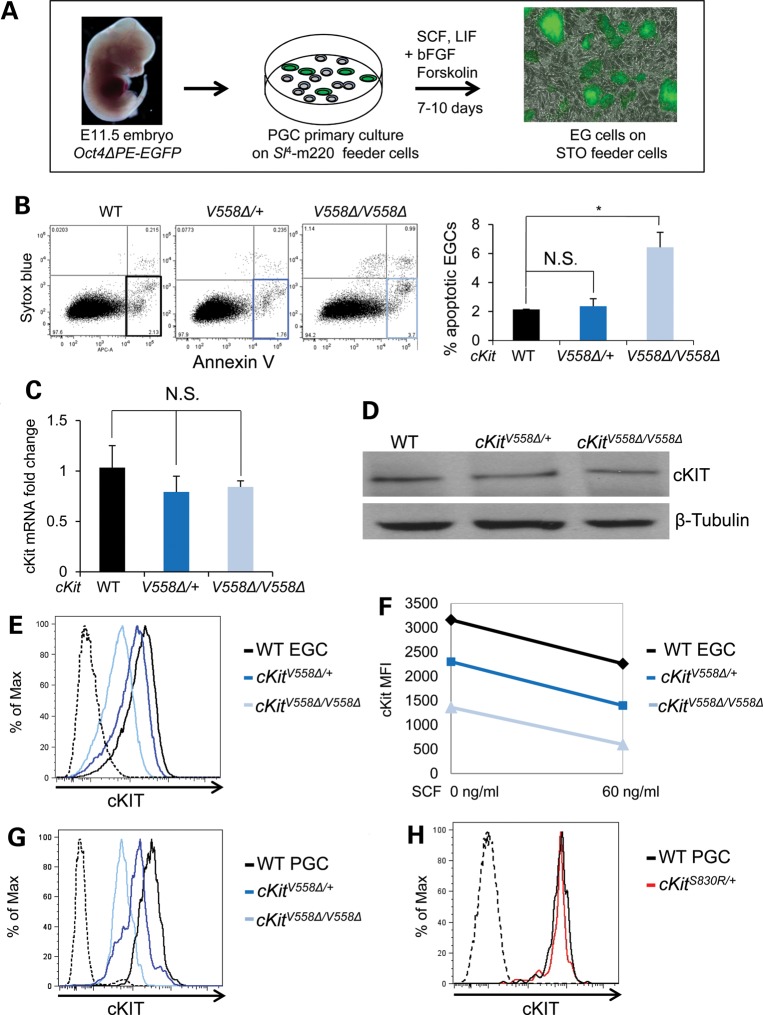
EGCs derived from cKitV558Δ PGCs recapitulate survival defects and display lower levels of surface cKIT. (A) EGC cell lines were derived from PGCs of E11.5 embryos harboring Oct4ΔPE:GFP as schematized and cultured on STO feeder cells (gray). (B) By annexin V staining, apoptosis was slightly increased in cKitV558Δ/V558Δ EGCs compared with WT or heterozygotes. (C) Similar levels of cKit transcript in all EGC genotypes were verified by qRT-PCR. (D) cKIT protein levels did not differ among all EGC genotypes by immunoblotting. (E–H) Live immunostaining with cKIT antibody revealed successively reduced surface expression on cKitV558Δ/+ and cKitV558Δ/V558Δ EGCs. SCF-induced downregulation of cKIT was comparable in all EGC genotypes (F). Similarly, cell surface cKIT reduction was observed in cKitV558Δ/+ and cKitV558Δ/V558Δ PGCs (G) but not on cKitS830R/+ PGCs (H). *P < 0.05; bars represent mean ± SEM.

