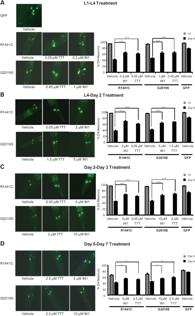
Figure 5.
Treatment with TTT-3002 and LRRK2-IN-1 arrests neurodegeneration in transgenic R1441C and G2019S worms. Transgenic C. elegans lines were treated with vehicle, TTT-3002 and LRRK2-IN-1 during L1–L4 stage (A), L4 to adult Day 2 (B), adult Day 2 to adult Day 3 (C) and adult Day 5 to adult Day 7 (D). When left untreated (vehicle control) and examined on adult Day 9, only ∼40% of ADE/CEP head DA neurons remained in both R1441C and G2019S transgenic C. elegans, while ∼80% of ADE/CEP head DA neurons were present in GFP control C. elegans. (A) Representative fluorescence images taken on adult Day 9 following treatments in liquid culture during L1–L4 stage with vehicle control, LRRK2-IN1 and TTT-3002 of transgenic C. elegans expressing GFP marker only or additionally R1441C- and G2019S-LRRK2 in DA neurons (left panel), and quantitation of DA neurons in the head region monitored at L4 stage (adult Day 0) and on adult Day 9 (right panel). (B) Representative fluorescence images taken on adult Day 9 following treatments in liquid culture from L4 to adult Day 2 with vehicle control, LRRK2-IN1 and TTT-3002 of transgenic C. elegans expressing GFP marker only or additionally R1441C- and G2019S-LRRK2 in DA neurons (left panel), and quantitation of DA neurons in the head region monitored at L4 stage (adult Day 0) and on adult Day 9 (right panel). (C) Representative fluorescence images taken on adult Day 9 following treatments in liquid culture from adult Day 2 to Day 3 with vehicle control, LRRK2-IN1 and TTT-3002 of transgenic C. elegans expressing GFP marker only or additionally R1441C- and G2019S-LRRK2 in DA neurons (left panel), and quantitation of DA neurons in the head region monitored at L4 stage (adult Day 0) and on adult Day 9 (right panel). (D) Representative fluorescence images taken on adult Day 9 following treatments in liquid culture from adult Day 5 to Day 7 with vehicle control, LRRK2-IN1 and TTT-3002 of transgenic C. elegans expressing GFP marker only or additionally R1441C- and G2019S-LRRK2 in DA neurons (left panel), and quantitation of DA neurons in the head region monitored on adult Day 5 and on adult Day 9 (right panel). In all the treatment regimens, both TTT-3002 and LRRK2-IN1 significantly enhanced the percentage of surviving DA neurons assayed on adult Day 9. DA neurons in the head region were monitored at the L4 stage, on adult Day 5 or adult Day 9 using a fluorescence microscope. Results were obtained from three independent experiments, each with about 30 worms per genotype for a given compound concentration. Values are expressed as mean ± SEM. ***P < 0.01 and *P < 0.05 by one-way ANOVA.

