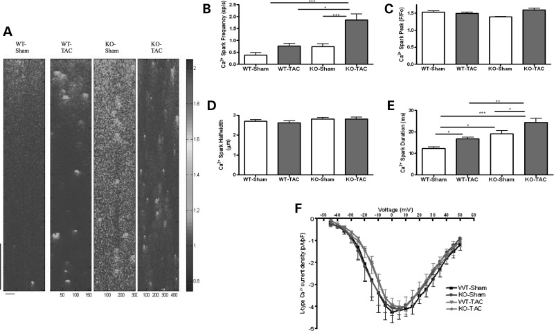
Figure 6.
Mechanical overload alters Ca2+ spark frequency in Tcap KO, but not WT and does not affect LTCC. (A) The figure shows line scans of quiescent ventricular cardiomyocytes. The colour chart is a normalized scale of fluorescence intensity (a marker of Ca2+ concentration). WT-Sham n = 34, WT-TAC n = 32, KO-Sham n = 31, KO-TAC n = 34. WT-Sham n = 11, WT-TAC n = 16, KO-Sham n = 13, KO-TAC n = 14. The vertical scale bar represents 768 ms and the horizontal scale bar represents 100 pixels. (B) The Ca2+ spark frequency increased only in KO, and Ca2+ peak and width did not change (C and D), whereas the Ca2+ spark duration increased more in KOs when compared with WT following TAC (E). (F) The L-type Ca2+ channel current–voltage relationship was unchanged.

