Figure 1.
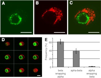
Human islet cells were isolated and cultured for 24 h. After a double immunofluorescence for insulin (red) and glucagon (green), islet cells were analyzed by confocal microscopy. A–C: Images showing a cell pair composed of one α-cell surrounded by one β-cell, the merged image is shown in panel C. The cell pair shown here is representative of cell pairs observed in different human cell preparations from at least 10 different pancreata. D: One series of consecutive merged images of a cell pair composed of one α-cell (green) surrounded by a β-cell (red). Scale bars, 10 μm. All heterologous contacts between α- and β-cells were coded according to their type of association: a β-cell wrapping an α-cell (β wrapping α), neutral apposition between α- and β- cells (α-β), and an α-cell wrapping a β-cell (α wrapping β). Results are shown as relative frequencies, and columns are means ± SEM of five islet cell preparations from five different pancreata. From all heterogenous contacts between α- and β-cells, the percentage of α-cells whose profile was round and the perimeter almost completely wrapped by a β-cell, as in panel D, was 38 ± 8 (means ± SEM of three experiments) (16). Reprinted with permission from the American Diabetes Association.

