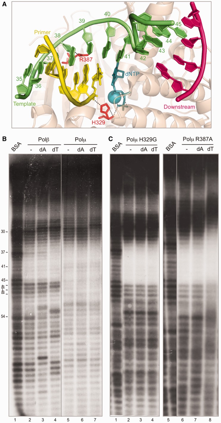
Figure 2.
Ternary complex formation in solution. (A) Structure of Polµ (shown in wheat-colored ribbons) ternary complex (2IHM), in which the DNA substrate and incoming nucleotide are shown in sticks with the following colors: dNTP, dark teal; template strand, green; primer strand, yellow; downstream strand, dark pink. Selected residues are shown in red sticks. Nucleotides in the template strand are numbered as in the footprinting assays for clarity. (B) Footprinting assay of the wild-type Polβ (1.5 µg) and Polµ (1.5 µg). When indicated, 100 µM dATP or dTTP were added, together with 2.5 mM MgCl2. The DNA substrate used, formed by hybridizing the oligonucleotides FP-T (template, labeled at its 5′-end), FP-P (primer) and FP-D (downstream) always contains a phosphate group at the 5′-end of the downstream strand. Gel was dried and the labeled fragments detected by autoradiography. (C) Footprinting assays of Polµ mutants H329G and R387K (1.5 µg) were carried out as described in (B).

