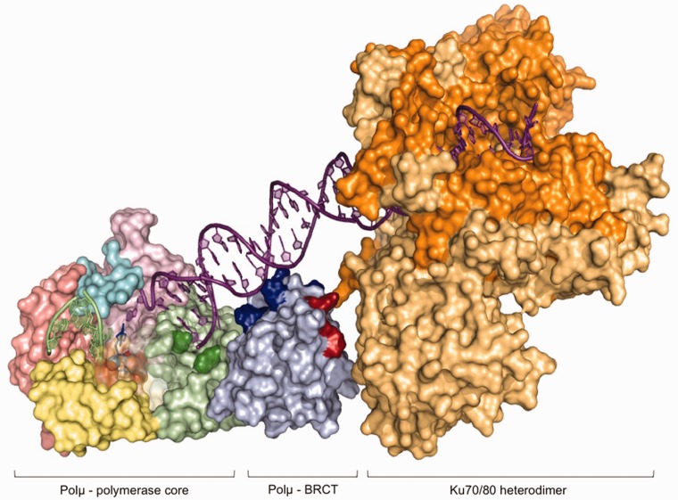
Figure 9.
Model of the interaction of Polµ with the Ku heterodimer and the DNA substrate through the BRCT domain. Computer-generated model of the predicted location of the Polµ β-like core, the BRCT domain and the Ku70/80 heterodimer on a single DNA end. The different domains in the Polµ core have been colored as follows: 8-kDa domain in green (with the residues forming the 5′-P pocket highlighted in dark green), the fingers subdomain in yellow, the palm subdomain in salmon, the thumb subdomain in pink and the Loop1 motif in cyan. The BRCT domain is colored light blue, with the residues predicted to be interacting with the DNA substrate in dark blue and the residues implicated in the interaction with the NHEJ factors in red. The two subunits of the Ku heterodimer are colored in dark and light orange.

