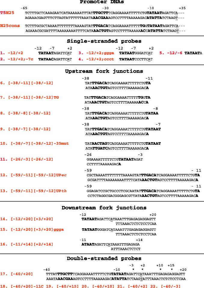
Figure 2.
Structures of DNA probes used. The probe names used in the text are in red. In fork junction probe names, numbers in left and right parentheses correspond to borders of upper and bottom strands of fork junctions with respect to the transcription start located at +1 (underlined). The −10 and −35 promoter element sequences are highlighted in larger size font. Asterisks above the [−40/+20] probe indicate positions where the fragments used in experiment shown in Figure 5 were truncated.

