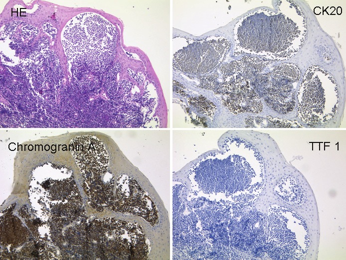
Figure 2.

The histological section of a resected specimen of the right pharyngeal tonsil shows infiltrates of a smallblue cell invasive tumour (A, HE). Tumour cells express the epithelial marker Cytokeratin 20 (B), the neuroendocrinemarker Chromogranin A (C) but not the neuroendocrine marker TTF1 (D) (all images x200). The tumour cells show thetypical immunohistochemical pattern of a Merkel cell tumour.
