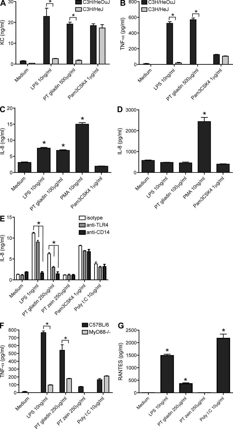
Figure 2.
Gliadin-induced innate immune responses are mediated via TLR4 and via the MyD88-dependent and -independent pathway. (A and B) KC (murine IL-8; A) and TNF (B) secretion in peritoneal macrophages isolated from TLR4-deficient C3H/HeJ mice compared with C3H/HOuJ wild-type mice stimulated with LPS or PT gliadin. The TLR2 agonist Pam3CSK4 served as cell viability control. (C and D) IL-8 secretion upon PT gliadin and LPS stimulation in 293 cells transfected with the TLR4–MD2–CD14 complex (C) and in untransfected cells (D). LPS and PMA served as positive controls, and the TLR2 agonist Pam3CSK4 served as negative control. (E) IL-8 secretion after preincubation with anti-TLR4 and anti-CD14 antibodies in monocyte-derived DC stimulated with PT gliadin and LPS. TLR2 agonist Pam3CSK4 and TLR3 agonist Poly I:C served as positive controls. (F) TNF secretion in peritoneal macrophages isolated from MyD88−/− mice compared with C57BL/6J wild-type mice upon PT gliadin stimulation. LPS served as positive control for the MyD88 knockdown, and TLR3 agonist Poly I:C served as cell viability control. (G) RANTES secretion in peritoneal macrophages isolated from C57BL/6J mice upon LPS, Poly I:C, and PT gliadin stimulation. *, P < 0.05 versus negative (positive) control (if not indicated otherwise); all graphs illustrate representative data from one of at least three independent experiments, and all experiments were performed in triplicates. Error bars depict standard errors of the mean.

