Figure 1.
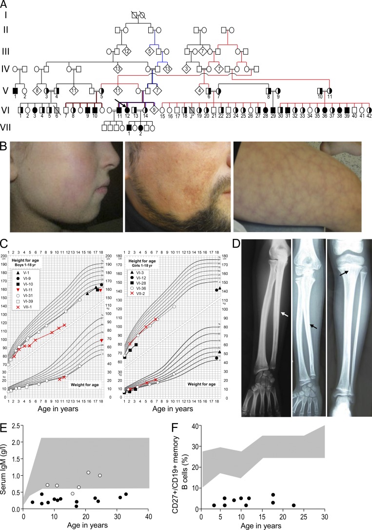
Pedigree and clinical manifestations in patients with the FILS phenotype. (A) Pedigree of the family with FILS. Family members having provided DNA are numbered. Completely closed symbols indicate affected individuals, and half closed symbols indicate a heterozygous carrier. An arrow designates the index case. Individuals clustered in a diamond symbol in generation IV and V were not analyzed. Gray symbols indicate patients that did not undergo genetic testing. Slashes indicate deceased persons, double horizontal lines consanguinity, and dotted lines presumed interrelations. Case J* died after a pulmonary infection at the age of 2 yr. (B) Facial profile photographs showing patient VI-36 at the age of 9, with livedo on the cheek and discrete malar hypoplasia (left), telangiectasia on the cheek of patient VI-11 at the age of 33 (middle) and, lastly, livedo on the thigh of patient VI-11 (right). (C) Growth charts for male (left) and female (right) patients, showing short stature in all instances and particularly substantial growth impairment in patients VI-3, VI-10, VI-11, VI-12, VI-28, and VII-1. (D) X ray of the forearm of case VI-3 (left) and patient VI-29 (middle) showing irregular diaphyseal hyperostosis (arrows) and x-ray of the leg of patient VI-29 (right) showing irregular diaphyseal hyperostosis and metaphyseal striations (arrow). (E) IgM levels in patients (closed dots) and heterozygous individuals (open dots) of different ages. The shaded area indicates our in-house normative values. (F) Percentages of memory B cells (CD27+/CD19+) in patients (closed dots). The shaded area indicates normal level from in-house control values.

