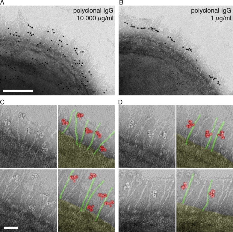
Figure 5.
Localization and orientation of polyclonal IgG at the bacterial surface. (A and B) Negative staining EM was used to visualize the localization and orientation of IgG bound to the bacterial surface. Gold-labeled IgG is found either widely scattered (A, 10,000 µg/ml IgG) or found along the midsection of the surface proteins (B, 1 µg/ml IgG) of wild-type S. pyogenes. Bar, 100 nm. Images are representative of two independent experiments. (C and D) High magnification shows single IgG molecules bound either via Fab (C, 10,000 µg/ml IgG) or via Fc (D, 1 µg/ml IgG). Two representative images with pseudocolor variants are shown of each experiment. Bar, 25 nm.

