Figure 1.
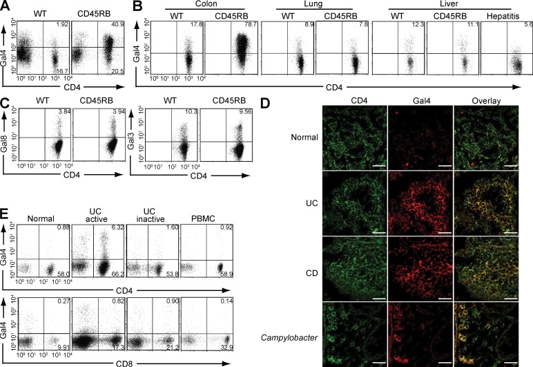
CAG. (A) Galectin-4 (Gal4) binding patterns on lamina propria cells (including CD4+ versus CD4− cells) in normal colon of WT mice and inflamed colon of CD45RB model are shown. Data shown are one representation of seven independent experiments. (B) Intensities of galectin-4 binding on CD4+ T cells from the colon, lung, and liver of WT mouse, CD45RB model, and concanavalin A–induced hepatitis model (right panel) are shown. The results are representative in 3–12 mice in each group. (C) Intensities of galectin-3 (Gal3) and galectin-8 (Gal8) binding on CD4+ T cells from the colon of WT mouse and from the inflamed colon of CD45RB model are shown. The results are representative in 3 mice in each group. (D) Colonic tissues from healthy individuals (n = 5) and from patients with UC (n = 6), CD (n = 4), or Campylobacter infection (n = 2) were co-stained with FITC–anti-CD4 (green, left) and Alexa Fluor 594–labeled recombinant human galectin-4 (red, middle). Overlap images are shown on the right. Bars, 25 µm. (E) Flow cytometric analysis shows the intensity of galectin-4 binding on CD4+ (top) or CD8+ (bottom) T cells from inflamed colon (active, n = 10), noninvolved colon (inactive, n = 5), and peripheral blood (PBMC, n = 2) of UC patients and from normal colon of control subjects (normal, n = 4).

