Figure 3.
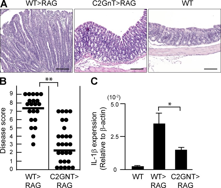
Pathogenic role of CAG in colitis of CD45RB model. CD4+CD45RBhigh naive T cells were purified from the spleen of WT (WT>RAG) versus T/C2GnT Tg (C2GnT>RAG) mice, and they were adoptively transferred i.v. into Rag1−/− mice. The recipients were sacrificed at 8 wk after transfer. (A) Representative colonic histologies (×10 objective) are shown. Colon of donor WT mouse is shown as normal control. Bars, 200 µm. (B) Disease scores evaluated by a combination of macroscopic and microscopic examinations are summarized. Each dot represents individual recipient. **, P < 0.0001. (C) Expression levels of IL-1β in the colonic tissues from WT mice and from Rag1−/− mice with transfer of WT-derived (WT>RAG) versus T/C2GnT tg–derived (C2GnT>RAG) CD4+ T cells are shown. n = 6/each group. *, P < 0.005.

