Figure 8.
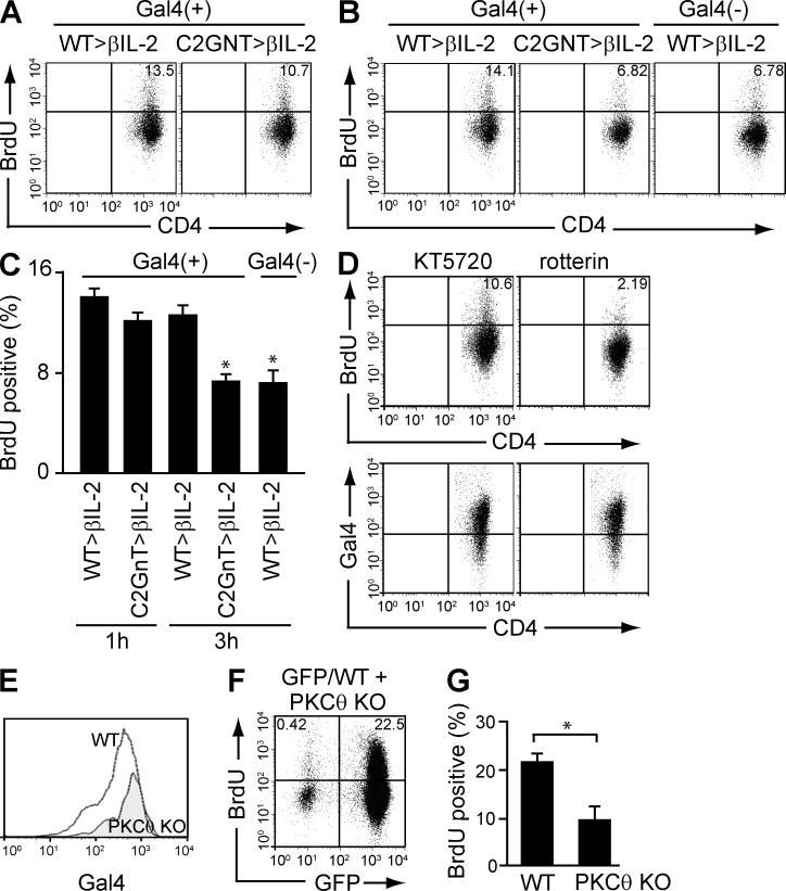
Requirement of PKCθ for CAG-mediated proliferation. (A-C) Purified colonic CD4+ T cells from βIL-2 DKO mice with reconstitution of WT-derived (WT>βIL2) versus T/C2GnT tg–derived (C2GnT>βIL2) memory CD4+ T cells were stimulated with anti-CD3/CD28–coated beads, and after removal of beads they were cultured again in the presence or absence of galectin-4 for 1 (A) and 3 h (B). They were pulsed with BrdU 1 h before the analysis and subjected to flow cytometric analysis. The percentage of BrdU+ cells is summarized in C. *, P < 0.01 (n = 4–7). (D) Purified colonic CD4+ T cells from βIL-2 DKO mice with reconstitution of WT-derived (WT>βIL2) versus T/C2GnT tg–derived (C2GnT>βIL2) memory CD4+ T cells were pretreated with KT5720 (left) or rotterin (right), and they were then stimulated with anti-CD3/CD28–coated beads. After removal of beads, they were cultured in the presence of galectin-4 for 3 h, with 1-h pulse with BrdU, and subjected to flow cytometric analysis for evaluation of BrdU incorporation (top) and galectin-4 binding (bottom). Data shown are one representation of two independent experiments. (E and F) Purified CD4+CD45RBlow memory T cells from the spleen of GFP Tg versus PKCθ−/− mice were co-transferred into βIL-2 DKO mice. The intensity of galectin-4 binding on (E) and the proliferative response of (F) reconstituted GFP+ CD4+ (PKCθ-intact) versus GFP− CD4+ (PKCθ-deficient) cells in the same recipient colon are shown. The mean (n = 4) of percentages of BrdU+ cells in GFP+ (PKCθ-intact) versus GFP− (PKCθ-deficient) CD4+ T cells are summarized in G. *, P < 0.01.

