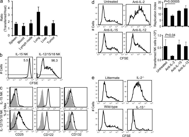
Figure 4.
Rapid proliferation of IL-12/15/18–preactivated NK cells in recipient mice is dependent on endogenous IL-2. NK cells (CD45.1+) were pretreated with IL-15 or IL-12/15/18 for 16 h and labeled with CFSE. RMA-S tumor–bearing mice received RT and 106 CFSE-labeled NK cells 3 h after irradiation as described in Fig. 2 b. (a) 4 d later, host and transferred cells were analyzed in the spleen, blood, lymph node, lung, liver, and tumor. The graph depicts the ratio between the numbers of transferred and host NK cells (mean + SD; n = 5). Data are representative of two independent experiments. (b) 4 d after transfer, in vivo proliferation of transferred NK cells was analyzed. Histograms were gated on CD3ε−NK1.1+CD45.1+ transferred NK cells, and one representative histogram from each group is shown (n = 5). Percentages of cells that proliferated are depicted. Data are representative of three independent experiments. (c) NK cells were treated with IL-15 or IL-12/15/18 for 16 h and stained with anti-CD25, -CD122, and -CD132 mAbs (bold black lines) or the isotype control (filled histograms). Data are representative of three independent experiments. (d) CFSE-labeled IL-12/15/18–pretreated NK cells (CD45.1+) were transferred into RMA-S tumor–bearing, irradiated mice (CD45.2+) as described in Fig. 2 b. Anti–IL-2, –IL-15, or –IL-12 mAb was applied as described in Materials and methods. 4 d after adoptive transfer, in vivo proliferation of transferred NK cells was analyzed. Cells were gated on CD3ε−NK1.1+CD45.1+ transferred NK cells, and one representative histogram from each group is shown (n = 3–5). The replication index indicating the fold expansion of the proliferating cells was calculated by FlowJo. Numbers of transferred NK cells in spleen are depicted. Graphs indicate mean + SD (n = 3–5). Data are representative of two independent experiments. (e) IL-2−/− mice or littermates (CD45.2+; top) and IL-15−/− or wild-type mice (CD45.2+; bottom) received RT and 106 CFSE-labeled preactivated NK cells (CD45.1+) 3 h after irradiation. 4 d later, CFSE dilution of transferred NK cells from spleen was determined. Data shown were gated on CD3ε−NK1.1+CD45.1+ transferred NK cells. One representative histogram from each group is shown (n = 3–5).

