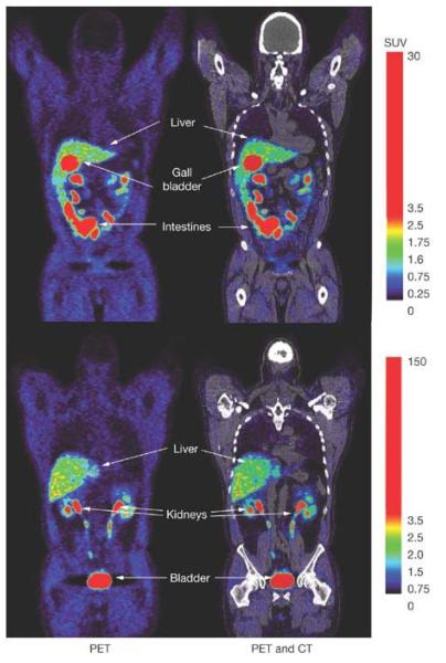
Figure 2.
Whole-body PET and PET/CT images of [18F]FHBG biodistribution in a human, two hours after it’s intravenous injection. Two coronal slices are shown to illustrate activity within the liver, gall-bladder, intestines, kidney’s and bladder, which are organs involved with [18F]FHBG’s clearance from the body. Background activity in all other tissues is relatively low, due to the absence of HSV1-tk or HSV1-sr39tk expressing cells within the body of this human volunteer.

