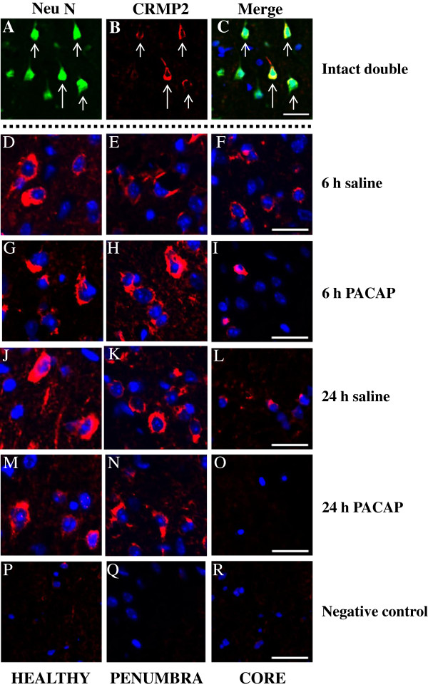
Figure 7.
Immunofluorescent staining. Neurons express CRMP2 at 6 and 24 h after permanent middle cerebral artery occlusion (PMCAO). A,B,C: triple immunofluorescence staining for CRMP2, NeuN and 4′, 6-diamidine-2-phenylindole dihydrochloride (DAPI); the merged picture displays expression of CRMP2 by NeuN positive neuron but not DAPI-positive neurons. D,G,J,M,P: healthy; E,H,K,N,Q: penumbra; and F,I,L,O,R: core. D-R: double immunofluorescence staining for CRMP2, and DAPI; the merged picture shows mutually exclusive expression of neuron. The merged picture shows membranous localization of CRMP2 surrounding DAPI-positive nuclei. All images were captured in the ipsilateral hippocampus. Intact (double): A, B, C; 6 h saline: D, E, F; 6-h pituitary adenylate cyclase-activating polypeptide (PACAP): G, H, I; 24 h saline: J, K, L; 24 h PACAP: M, N, O; negative control: P, Q, R. Green - NeuN, Red - CRMP2, and Blue - DAPI. Scale bar: 20 μm. The sections were prepared as described in Methods; see also Additional file 8: Figure S6.

