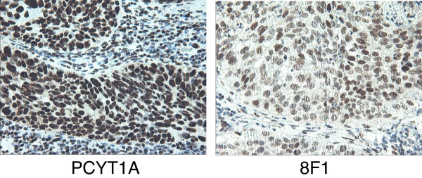
Figure 4.
The immunohistochemistry staining on NSCLC tissue sections with rabbit monoclonal anti-PCYT1A antibody or 8F1 mouse monoclonal anti-ERCC1 antibody. Pathologist-validated NSCLC FFPE tissue blocks were cut into thin sections, and then further deparaffinized, re-hydrated before IHC testing. The primary antibodies (Left: rabbit anti-PCYT1A mAb, Right: 8F1 anti-ERCC1 mAb) used for these experiments were diluted at 1:150 dilution with blocking buffer.

