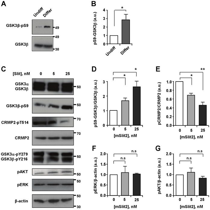
Figure 6. mSlit2 induces phosphorylation and inactivation of GSK3β.
(A, B) Undifferentiated and differentiated CAD cell lysates were subjected to Western blot analysis to compare the level of phospho-GSK3β. Lysates for differentiated CAD cells were collected at 24 hr after the induction of differentiation. For quantification in (B), the level of phospho-GSK3β was normalized against GAPDH loading control. Presented are representative immunoblots (A) and quantification of Western blot analysis (B). (C–G) Differentiated CAD cells were stimulated with two different concentrations of recombinant mSlit2 (5 nM or 25 nM). Protein lysates were prepared and subjected to Western blot analysis. Expression and/or activation/inactivation of GSK3 as well as other kinases, such as AKT and ERK1/2, were emxamined. The level of phosphorylated CRMP2, a substrate of GSK3 was also monitored. Representative immunoblots are shown in (C) and quantification of Western blot analysis is shown in (D–G). * p<0.05; ** p<0.01; n.s. not statistically significant.

