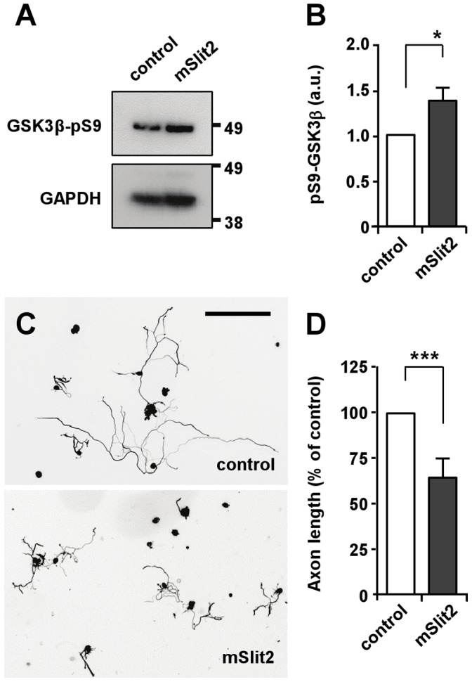
Figure 8. mSlit2 inhibits GSK3β and neurite outgrowth in DRG neurons.

(A, B) DRG neurons from conditioning lesioned mice were treated with recombinant mSlit2 (25 nM) or vehicle control. Protein lysates were subjected to Western blot analysis and the level of phospho-GSK3β was examined. For quantification in (B), the level of phospho-GSK3β was normalized against GAPDH loading control. Presented are representative immunoblots (A) and quantification of Western blot analysis (B). * p<0.05. (C, D) DRG neurons from conditioning lesioned mice were cultured overnight in the presence or absence of mSlit2 (25 nM). Neurons were then fixed and stained for βIII-tubulin to measure neurite length. Presented are representative images (C) and quantification of neurite length (D). Bar, 200 µm. *** p<0.001.
