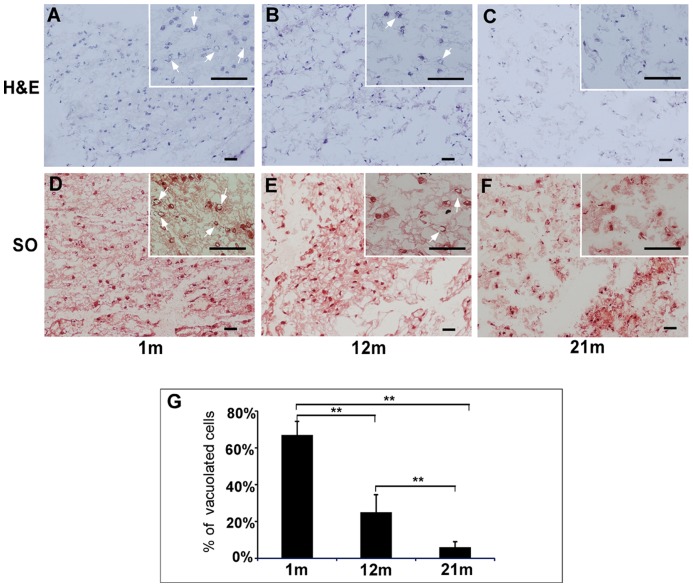
Figure 1. Histological characterization of NP tissue from IVD of rats at different ages.
Frozen tissue sections from (A, D) 1 m, (B, E) 12 m and (C, F) 21 m-old rats were stained with (A, B, C) Hematoxylin and Eosin (H&E) for cell structure and (D, E, F) Safranin O (SO) for proteoglycan content. Representative images are shown and the inserts of small images shown higher magnifications of interested regions. Bar: 50 µm. m: month. Arrows in subfigures indicate vacuolated morphology. (G) The percentage of cells with vacuolated morphology indicates an age-dependent change in NP tissue. **p<0.01 as compared to 1 m NP (one-factor ANOVA, n = 8). (TIF)

