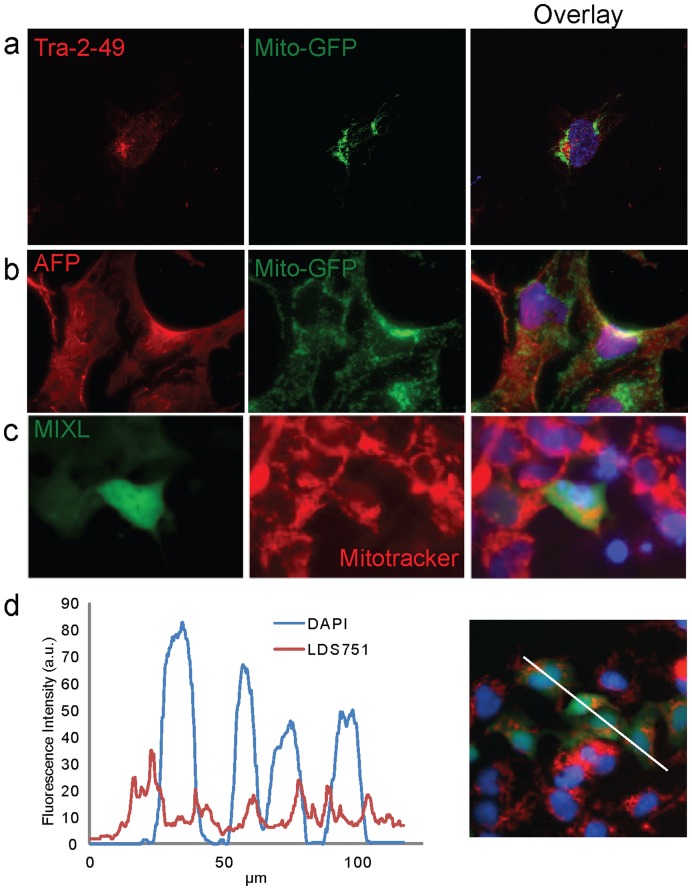
Figure 5. Variable mitochondrial localisation during lineage specific differentiation.
(a) Mitochondria in hESC are localised near the nucleus. (b) Mitochondria in AFP positive endoderm lineage cells. Mitochondria in AFP positive cells display a granular, dispersed localisation through the whole cell. (c and d) Mitochondria in MIXL1 positive cells (Mesendoderm) display a densely packed, perinuclear localisation based on MitoTracker far red (c) and LDS-751 (d) staining. AFP, alpha fetoprotein. Images (a-c) are 150 µm wide. Line profile in (d) represents 120 µm.

