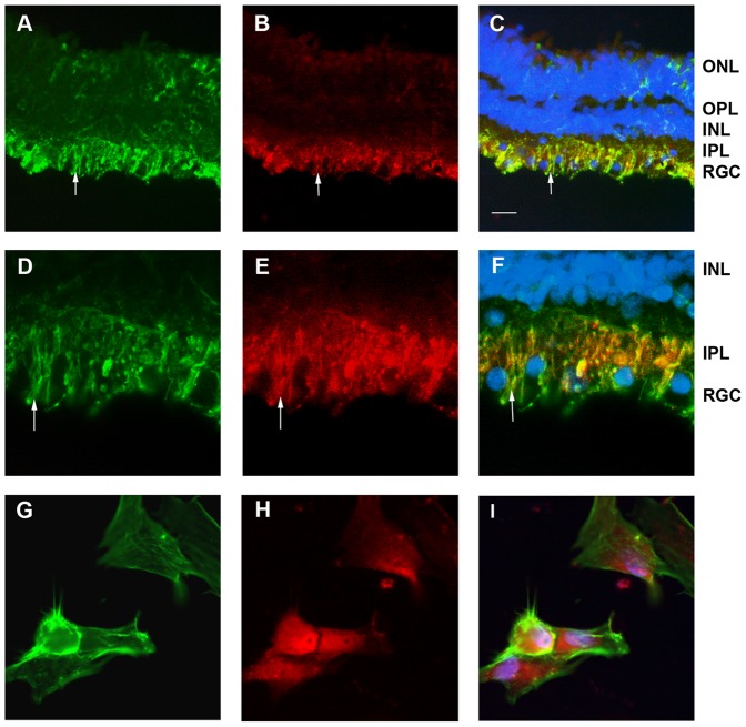
Figure 2. hCG-β localization in human retina.
Vimentin labeled Muller glial cell of the retina mainly in the GCL and IPL (A, C, D, F). Müller glia cell processes are labeled with vimentin (A, D) and hCG-β (B, E) between the ganglion nucleus that are labeled in blue in the GCL (C, F). Arrow (A–F) depicts glia cell endfeet that is double labeled with hCG-β (C, F). Diffuse staining against hCG-β suggests a secretion of hCG in the retina (B, E). hCG-β localization in human RPE. Primary RPE cells kept in culture were stained with phalloidin (G) and hCG-β (H), Merged image (I) revealed the expression of this gonadotropin in the RPE cells. Scale bar is 30 µm for A–C 15 µm for D–F and 10 µm for G–I. A, D: Mouse monoclonal anti vimentin (Abcam ref# ab28028) use 1/100 in PBS, 0.1% BSA, 0.01% triton x100; B, E, H, I: Rabbit polyclonal anti hCG-β (Abcam ref# ab9376) use 1/100 in PBS, 0.1% BSA, 0.01% triton x100; G, I : Phalloidin-488 (Millipore ref# A12379) use at 1/50 in PBS; C, F, I : Merged image, with blue channel corresponding to DAPI nucleus staining used at 1/1000. ONL = Outer nuclear layer, OPL = outer plexiform layer, INL = Inner nuclear layer, IPL = Inner plexiform layer, GCL = Ganglion cell layer.

