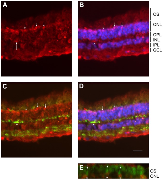
Figure 3. LHR localization in human retina.
LHR was found expressed throughout the human retina with an enhanced signal in the photoreceptor layer (ONL) especially in the most superficial layer (small arrows in A–D), signal was found around cell soma in the ONL but also rarely in the OPL (long arrow in A–C) and in the INL. In addition a diffuse signal was found in the basal part of the GCL. The DAPI co-staining confirmed that photoreceptor in the most outer layer were labeled (small arrows in B). Synaptophysin labeled presynaptic structures in the OPL and IPL synaptic layers (C, D). Large stained structures in the OPL corresponded to a cone terminals (long arrow in C) that was also found labeled with LHR antibody (long arrow in A–C) suggesting that stained neurons in the ONL with anti-LHR may correspond to cone photoreceptors. Photoreceptor labeled with LHR antibody in the most superficial layer of the ONL (asterisks in E) exhibit outer segment with a cone shape as stained with anti CD133 antibody (small arrow in E) thus confirming the cone identity of the LHR-positive photoreceptors. Scale bar is 30 µm for A–C and 15 µm for E. OS = outer segment, ONL = outer nuclear layer, OPL = outer plexiform layer, INL = inner nuclear layer, IPL = inner plexiform layer, GCL = ganglion cell layer. A–E : Red channel, rabbit polyclonal anti LHR (Santa Cruz biotechnology ref# ab25828) use 1/100 in PBS, 0.1% BSA, 0.01% triton x100; B : Merged image, with blue channel corresponding to DAPI nucleus staining used at 1/1000; C : Merged image, with synaptophysin (green channel) using mouse monoclonal anti synaptophysin (Millipore ref# MAB368) at1/100 in PBS, 0.1% BSA, 0.01% triton x100; D: Merged image with synaptophysin (green) and DAPI (blue); E : Merged image, with green channel corresponding to CD133 staining using mouse monoclonal anti CD133 (eBioscience ref #14–1331) at 1/1000in PBS, 0.1% BSA, 0.01% triton x100.

