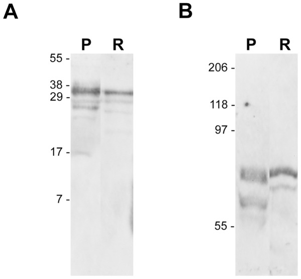
Figure 5. Western blot analysis of hCG and its receptor in retina. A).

Both placental and retinal extracts expressed hCG as detected on western blot using an antibody recognizing the whole hormone (Abcam ab 54410). Using 0.1 and 30 µg of protein extract from placenta (P) and Retina (R) respectively a major band is found in both tissues at an apparent molecular weight of 37 kDa corresponding to the heterodimer predicted sized. Smaller bands are seen in the placenta (17, 27, 29 kDa) and in the retina (20, 27, 29 kDa). B) LHR was detected in 10 µg of placental and retinal extract loaded on a 7.5% SDS gel. Major upper band at 85 kDa corresponds to the glycosylated form of the receptor whereas lower bands observes in placenta at 70 kDa (doublet) and in retina at 80 kDa may account for partially or un-glycosylated products.
