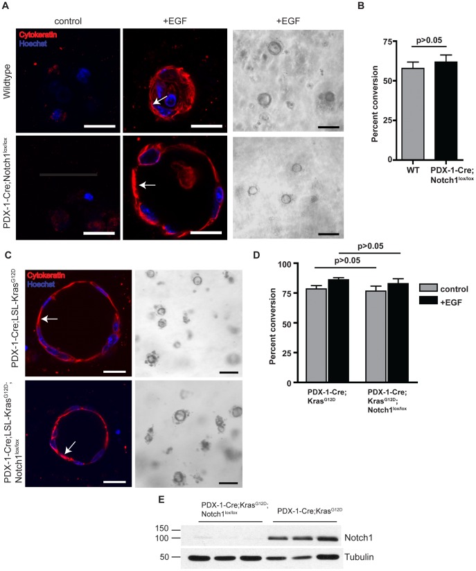
Figure 1. Notch1 is not required for oncogenic K-ras mediated ADM in vitro.
(A) Pancreatic explants from wildtype and PDX-1-Cre;Notch1lox/lox mice embedded in collagen either untreated (control) or treated with EGF (20 µg/mL). Cells are immunostained for expression of pan-cytokeratin (red) at day 5. Nuclei are stained with Hoechst dye (blue). Scale bar, 20 µm. Arrows indicate cytokeratin-positive ductal cells. Representative brightfield images are shown at day 5 in the presence of EGF. Scale bar, 100 µm. (B) Quantitative analysis of percent ductal cyst conversion on day 5 in explants isolated from wildtype and PDX-1-Cre;Notch1lox/lox mice. n = 3 for each group. (C) Pancreatic explants from PDX-1-Cre;LSL-KrasG12D and PDX-1-Cre;LSL-KrasG12D;Notch1lox/lox mice were isolated at day 2 in the absence of EGF. Cells are immunostained for pan-cytokeratin (red) and Hoechst dye (blue). Scale bar, 20 µm. Arrows indicate cytokeratin-positive ductal cells. Representative brightfield images of cyst formation are shown. Scale bar, 100 µm. (D) Quantitative analysis of percent ductal cyst conversion at day 2 in explants isolated from PDX-1-Cre;LSL-KrasG12D (n = 5) and PDX-1-Cre;LSL-KrasG12D;Notch1lox/lox mice (n = 6). (E) Western blot analysis of Notch1 expression in acinar cells isolated from PDX-1-Cre;LSL-KrasG12D and PDX-1-Cre;LSL-KrasG12D;Notch1lox/lox mice; tubulin as loading control. Three samples are shown for each genotype.

