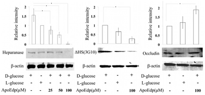
Figure 1. The protective effect of apoEdp on hRECs injured by high glucose in the cell medium.
(A). The hRECs were treated with varying doses of 25, 50, and 100 µM of apoEdp in the presence of 30 mM D-glucose or L-glucose (osmotic control). Western blot analysis of proteins extracted from high sugar-injured cells following 72 hours of apoEdp treatment revealed dose-dependent inhibition of heparanase expression. The 100 µM apoEdp treatment inhibited (*P≤0.05) heparanase expression to the lowest level of the 3 treatment groups assessed. (B). To detect the effect of hyperglycemia-induced HS loss (Δ-HS) in hRECs treated with 100 µM of apoEdp, a commercial monoclonal antibody (3G10) specific to neoepitope generated by heparanase digestion of HSPG was used. A 100 µM apoEdp treatment significantly inhibited (*P≤0.05) the loss of Δ-HS in hRECs after 72 hours of high glucose treatment compared to mock treatment (high glucose but no apoEdp treatment). (C). ApoEdp (100 µM) treatment prevented the loss of tight junction protein occludin in hRECs under hyperglycemic conditions. The hRECs were incubated for 72 hours with or without apoEdp (100 µM) in the presence of hyperglycemia and assayed for the expression of occludin. Each value represents the mean ± SE of results in three independent experiments (*P≤0.05). β-actin served as the loading control.

