
Figure 3. ApoEdp treatment restores retinal ZO-1 protein in diabetic mice as determined by immunohistochemistry.
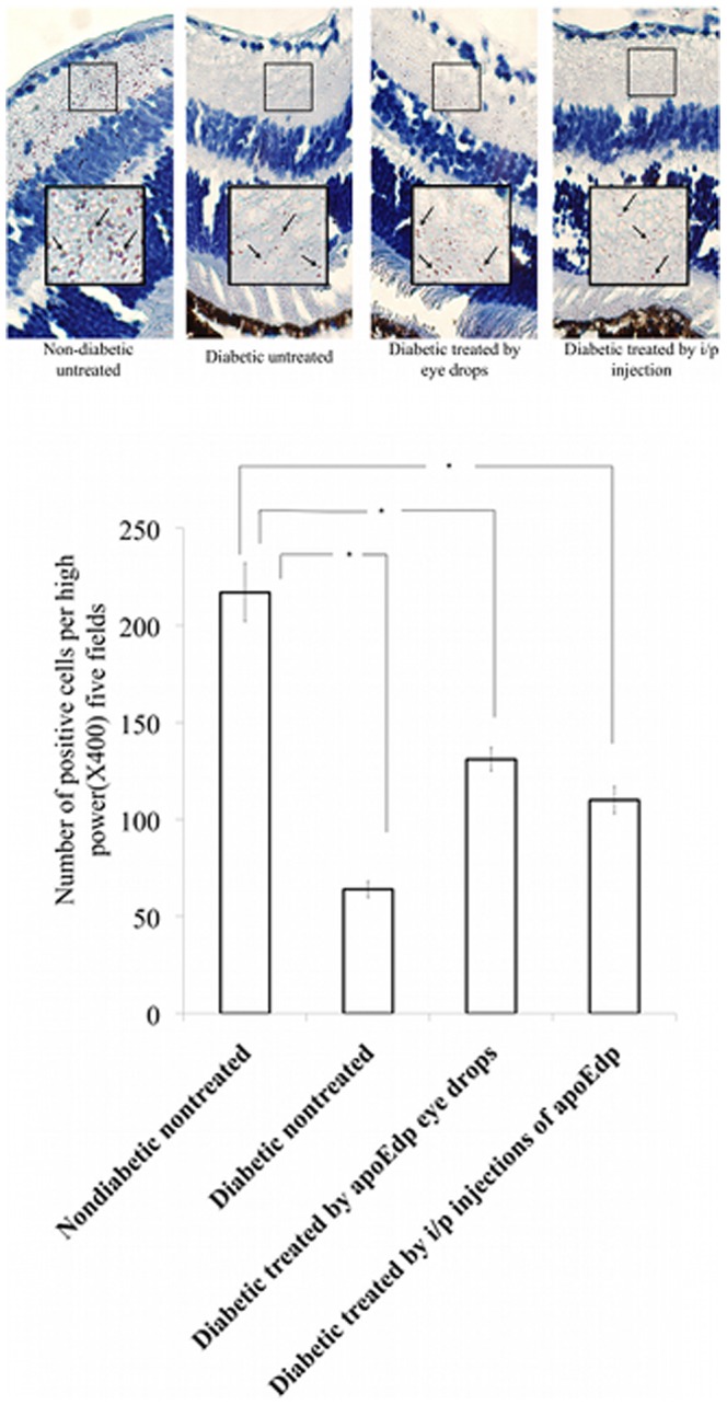
(A). Immunohistochemical analysis of formalin-fixed sections of mouse retinas were stained with zona occludin-1(ZO-1) specific antibody. Mean ±SEM of ZO-1 positive cells in five high power (×400) fields were counted. The arrows in the four photos indicate a ZO-1 positive cell. (B): The immunohistochemical quantitative analysis of ZO-1 protein in mouse retinas. Four treatment groups were used. The number of positive cells per high-power field is determined and the (mean ±SEM) of the ZO-1 positive cells are provided. The significant difference was detected in the ZO-1 cells between the diabetic, untreated eye, and the two groups of apoEdp treated mice retinas. Treatment with either topical 1% apoEdp drops or intraperitoneal injections of apoEdp for 14 consecutive days significantly improved the relationship of the number of increased positive cells compared to the untreated, diabetic retina.