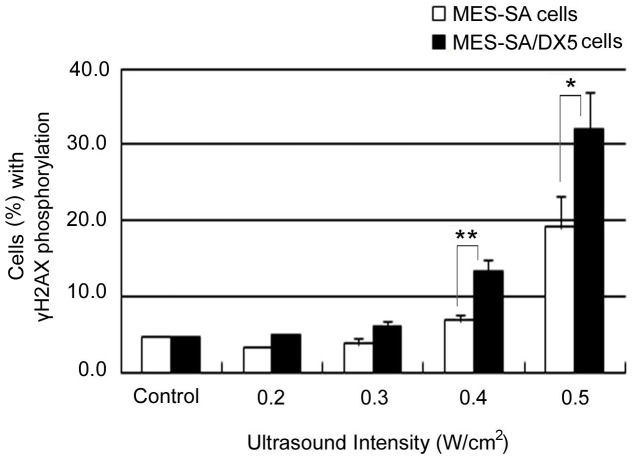
Figure 4. The extent of histone H2AX phosphorylation in MES-SA and MES-SA/DX5.
Cells were fixed 15 min after exposure to ultrasound at different intensities. Cells were assayed flow cytometrically. Data points are presented as mean ± SEM. Asterisks (*) denote the statistical significance between MES-SA and MES-SA/DX5 at respective intensities.

