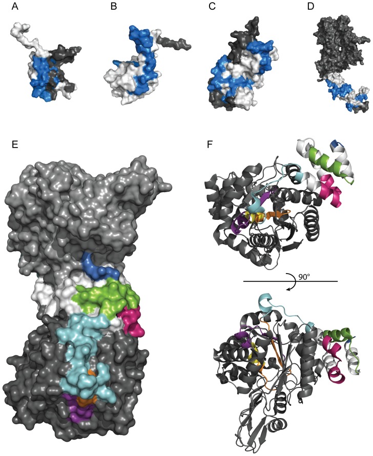
Figure 6. The three-dimensional structure of the epitopes.
Crystal or solution structures of antigens with highlighted antigenic determinants common in all three immunizations determined by bead array mapping in blue. The molecular surface of the protein is colored grey and the antigen part is highlighted in white. The images were acquired using MacPyMOL. Structures of (A) RRM domain of HNRNPH2 (1wez.pdb), (B) PDZ domain of SYNJ2BP (2eno.pdb), (C) PDXP (2cfs.pdb). (D) extracellular domain of ERBB2 (1n8z.pdb). (E) Crystal structure of homodimer TYMP (2jof.pdb) showing the molecular surface with indicated epitopes (blue, green, pink, cyan, orange, yellow and purple) used for affinity purification of monospecific antibodies. (F), View of monomeric TYMP showing secondary structural features of epitopes.

