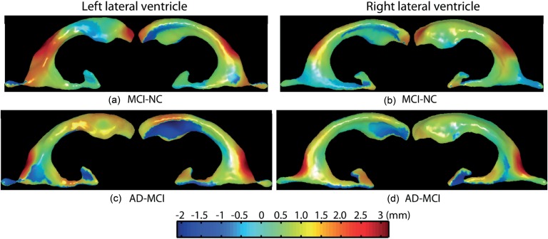
Figure 3. Shape differences of the lateral ventricles among normal controls (NC), mild cognitive impairment (MCI), and Alzheimer's Disease (AD).
Panels (a,b) respectively show group differences in the left and right lateral ventricular surface deformations between MCI and NC. Panels (c,d) respectively show group differences in the left and right lateral ventricular surface deformations between AD and MCI. Warm color denotes regions where structures have surface outward-deformation in the former group when compared with the latter group, while cool color denotes regions where structures have surface inward-deformation in the former group when compared with the latter group.

